肝臓のセグメンテーション
肝臓セグメンテーションは、肝臓がんの支援診断における重要な前処理である。本研究では、深層学習を用いた肝臓セグメンテーション法を数多く開発した。画像セグメンテーションに広く用いられるU-Netを改良したU-Net3+ (Fig.1)や、医用解剖アトラスを先験情報とするDeep Atlas Prior法(Fig.2)などを開発した。肝臓のセグメンテーション精度は95%に達した。それらの成果は信号処理、医用画像解析分野のトップコンファレンスIEEE ICASSP, MICCAIなどで発表した。
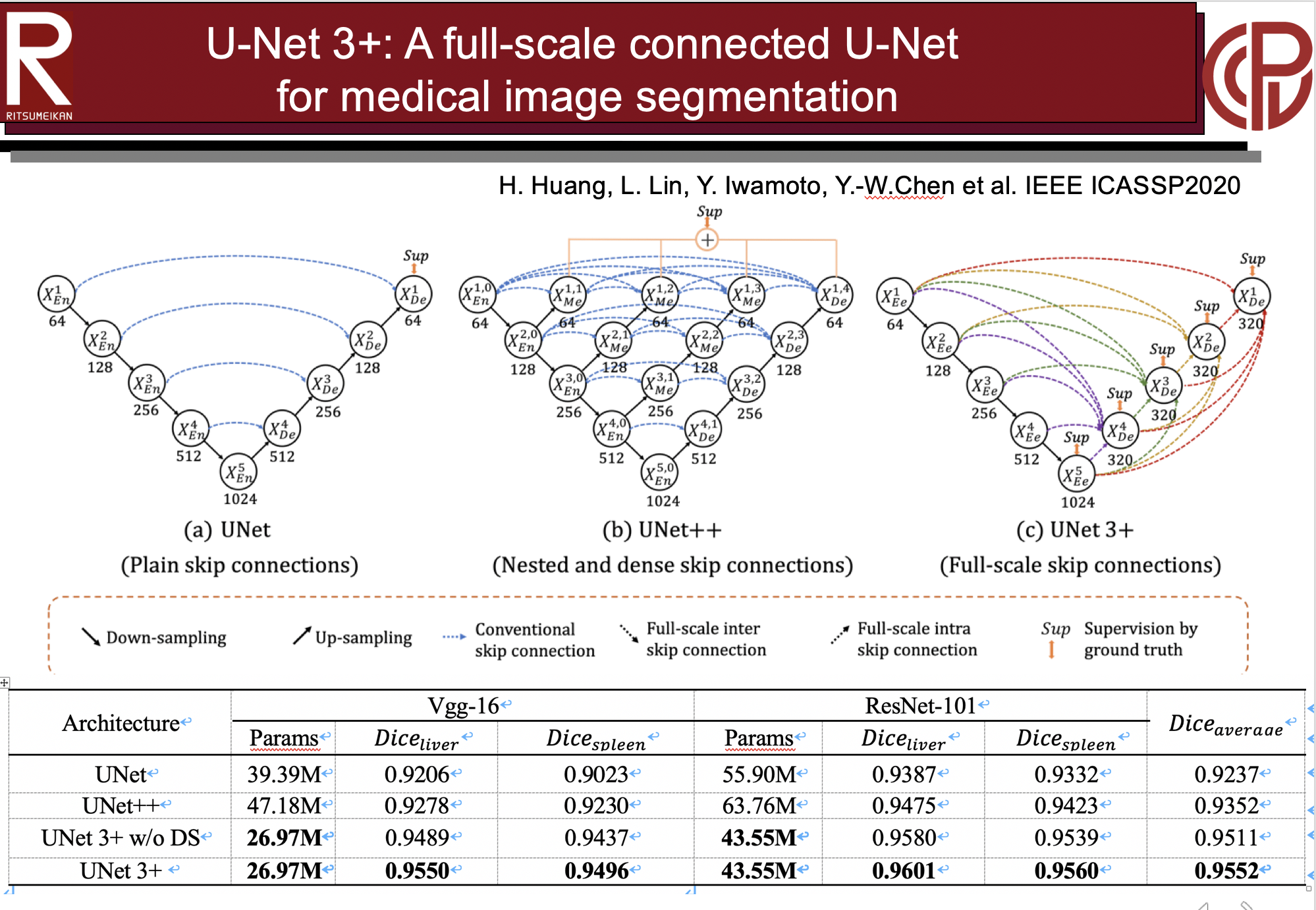
Fig.1. 提案法U-Net3+(c)は既存法のU-Net(a)とU-Net++(b)に比べ、Full-scal connection構造となっているので、高精度な肝臓セグメンテーションが実現できた。成果はIEEE ICASSP2020にて発表。
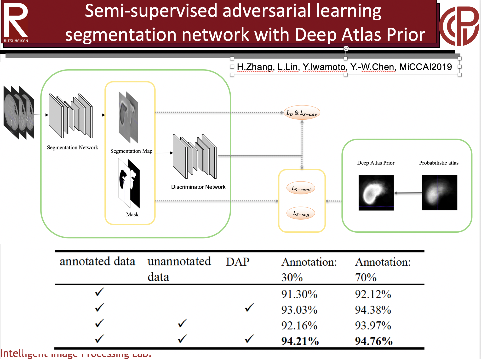
Fig.2. 医用解剖アトラスを先験情報とするDeep Atlas PriorとGANを組み合わせたSemi-supervised肝臓セグメンテーション法。少数なラベル付き学習データでも高精度な肝臓セグメンテーションができた。成果はMICCAI2019にて発表。
1-1. 医用画像データベースの構築
関連発表論文:
1. Huiming Huang, Lanfen Lin, Ruofeng Tong, Hongjie Hu, Qiaowei Zhang, Yutaro Iwamoto, Xian-Hua Han, Yen-Wei Chen, Jian Wu, ”UNET 3+: A Full-Scale Connected UNET for Medical Image Segmentation,” Proc. of the 45th IEEE International Conference on Acoustics, Speech, and Signal Processing (IEEE ICASSP2020), pp.1055-1059, Barcelona, Spain, May 4-8, 2020. (Oral)(Link)
2. Han Zheng, Lanfen Lin, Hongjie Hu, Qiaowei Zhang, Qingqing Chen, Yutaro Iwamoto, Xianhua Han, Yen-Wei Chen, Ruofeng Tong, Jian Wu, “Semi-supervised Segmentation of Liver Using Adversarial Learning with Deep Atlas Prior,” In: Shen D. et al. (eds) Medical Image Computing and Computer Assisted Intervention – MICCAI 2019. Lecture Notes in Computer Science, LNCS11769, Springer, pp.148-156, 2019. (Link)
3. Kawahara Toshiki, Yinhao Li and Yutaro Iwamoto, Lanfen Lin, Yen-Wei Chen, “A Lightweight Deep Network for 3D Medical Image Segmentation,” Proc. of 2020 IEEE 8th Global Conference on Consumer Electronics (GCCE 2020), Kobe, Japan, Oct.12-14, 2020(Link)
4. Hikari Jinbo; Titinunt Kitrungrotsaku; Yutaro Iwamoto; Lanfen Lin; Hongjie Hu, Yen-Wei Chen, “Development of an Interactive Semantic Medical Image Segmentation System,” Proc. of 2020 IEEE 8th Global Conference on Consumer Electronics (GCCE 2020), Kobe, Japan, Oct.12-14, 2020 (Link)
5. Ye Yuan, Yen-Wei Chen, Chunhua Dong, Hai Yu and *Zhiliang Zhu: “Hybrid Method Combining Superpixel, Random Walk and Active Contour Model for Fast and Accurate Liver Segmentation,” Computeried Medical Imaging and Graphics, Vol.70, pp. 119-134, 2018 (Impact factor: 3.750).(Link)
6. Chunhua Dong, Yen-Wei Chen, Lanfen Lin, Hongjie Hu, Chongwu Jin, Huajun Yu, Tomoko Tateyama, Xian-hua Han, “Simultaneous Segmentation of Multiple Organs Using Random Walks,” Journal of Information Processing Society of Japan, Vol.24, No.2, pp.320-329(2016). (Link)
7. Chunhua Dong, *Yen-Wei Chen, Amir Hossein Foruzan, Lanfen Lin, Xian-hua Han, Tomoko Tateyama, Xing Wu, Gang Xu and Huiyan Jiang, “Segmentation of liver and spleen based on computational anatomy models,” Computers in Biology and Medicine, Vol. 67, pp.146-160, 2015 (Impact factor: 3.434).(Link)
8. Titinunt Kitrungrotsakul, Chunhua Dong, Tomoko Tateyama, Xian-Hua Han, Yen-Wei Chen,”Interactive Segmentation and Visualization System for Medical Images on Mobile Devices,” J. Adv. Simulat. Sci Eng., Vol.2, No.1, pp.96-107, 2015 (Link)
9. A. H. Foruzan, Y.-W. Chen at. al., “Segmentation of Liver in Low-contrast Images Using K-Means Clustering and Geodesic Active Contour Algorithms,” IEICE Trans., Vol.E96-D, pp.798-807, 2013 (Impact factor: 0.449). (Link)
1-3. 肝臓腫瘍の検出とセグメンテーション
1-4. 肝臓腫瘍の鑑別
1-5. 肝臓がんの早期再発予測
1-6. 肝臓腫瘍の類似症例検索